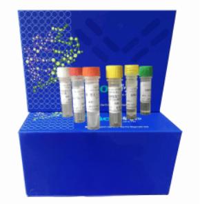
Gαo Activation Assay Kit
 产品名称 活性试剂盒 产品介绍
Configuration-specific Monoclonal Antibody Based
Gαo Activation Assay Kit
Catalog Number:CR-W80901
20 assays
Product Description
A structurally diverse repertoire of ligands, from photons to large peptides, activates G protein-coupled
receptors (GPCRs) to elicit their physiological functions. Ligand-bound GPCRs, in turn, function as
guanine nucleotide exchange factors catalyzing the exchange of GDP bound on the Gα subunit with
GTP in the presence of Gβγ, causing the dissociation of the Gα subunit from the Gβγ dimer to form
two functional units (Gα and Gβγ). Both Gα and Gβγ subunits signal to various cellular signaling
pathways. Based on the sequence and functional homologies, G proteins are grouped into four families:
Gs, Gi, Gq, and G12.
Gαi family (including Gαo) is the largest family of G proteins. They relay signals from many GPCRs
to regualte various biological functions. There were no direct methods to measure the activation of
Gαo proteins by receptors (until this assay kit). Most reports used one of the downstream pathway, i.e.
the inhibition of adenylyl cyclases, as a readout.
Bioyears Biosciences Gαo Activation Assay Kit provides a direct measurement of the activation of Gαo
proteins. This is a simple and fast tool to monitor the activation of Gαo. Each kit provides sufficient
quantities to perform 20 assays.
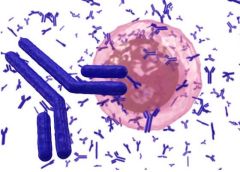
Bioyears Biosciences Gαo Activation Assay Kit is based on the monoclonal antibody specifically
recognizing the active GTP-bound Gαo proteins. This monoclonal antibody has much lower affinity
towards the inactive Gαo proteins. Therefore, after activation by receptor signals, active GTP-bound
Gαo proteins could be immunoprecipitated by this monoclonal antibody and further quantified by
western blot with another anti Gαo antibody.
Assay Principle
Bioyearst Biosciences Gαo Activation Assay Kit is an immunoprecipitation/western blot assay to
measure the levels of active GTP-bound Gαo proteins, either from cell extracts or from in vitro GTPγS
loaded Gαo proteins. Briefly, the anti active Gαo monoclonal antibody will specifically bind to active
Gαo protein. This antibody/Gαo complex will then be pulled down by protein A/G agarose. The
precipitated active Gαo proteins will be detected by immunoblots with another anti Gαo antibody.
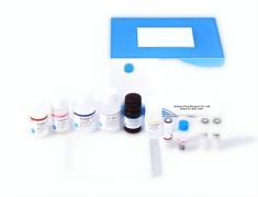
Kit Components
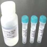
1. Anti active Gαo, Mouse Monoclonal Antibody (Catalog No. CR-M26907): One vial – 22 µL (1mg/mL) in
PBS, pH 7.4, contained 50% glycerol. This antibody specifically recognizes GTP- Gαo from all ertebrates.
2. Protein A/G Agarose (Catalog No. 30301): One vial – 400 µL of 50% slurry.
3. 5X Assay/Lysis Buffer (Catalog No. 30303): One bottle – 30 mL of 250 mM Tris-HCl, pH 7.4,750 mM
NaCl, 5 mM EDTA, 5% Triton X-100.
4. Anti Gα0, Rabbit Polyclonal Antibody (Catalog No. CR-R21015): One vial – 22 µL(1 mg/mL) in PBS, pH 7.4,
contained 50% glycerol.
5. 100 X GTPγS (Catalog No. 30302): One vial –100 µL at 10 mM, use 5 µL of GTPγS for GTP-labeling of
0.5 mL of cell lysate.
6. 100 X GDP (Catalog No. 30304): One vial –100 µL at 100 mM, use 5 µL of GDP for GDP-labeling of 0.5
mL of cell lysate.
Storage
Store all kit components at 4ºC until their expiration dates. For long-term usage (more than 6 months),
please keep the antibodies at -20oC.
Materials Needed but Not Supplied
1. Stimulated and non-stimulated cell lysates
2. Protease inhibitors
3. 4 °C tube rocker or shaker
4. 1 M MgCl2
5. 2X reducing SDS-PAGE sample buffer
6. Electrophoresis and immunoblotting systems
7. Immunoblotting wash buffer such as TBST (10 mM Tris-HCl, pH 7.4, 0.15 M NaCl, 0.05 %Tween-20)
8. Immunoblotting blocking buffer (TBST containing 5 % Non-fat Dry Milk or 3 % BSA)
9. PVDF or nitrocellulose membrane
10. Secondary Antibody
11. ECL Detection Reagents
Reagent Preparation
• 1X Assay/Lysis Buffer: Mix the 5X Stock briefly and dilute to 1X in deionized water. Just prior to
usage, add protease inhibitors such as 1 mM PMSF, 10 µg/mL leupeptin, and 10 µg/mL aprotinin.
Sample Preparation
Adherent Cells
1. Culture cells (one 10-cm plate, ~ 107cells) to approximately 80-90 % confluence. Stimulate cells with
activator or inhibitor as desired.
2. Aspirate the culture media and wash twice with ice-cold PBS.
3. Completely remove the final PBS wash and add ice-cold 1X Assay/Lysis Buffer to the cells (0.5- 1 mL per
10 cm tissue culture plate).
4. Place the culture plates on ice for 10-20 minutes.
5. Detach the cells from the plates by scraping with a cell scraper.
6. Transfer the lysates to appropriate size tubes and place on ice.
7. If nuclear lysis occurs, the cell lysates may become very viscous and difficult to pipette. If this occurs,
lysates can be passed through a 27½-gauge syringe needle 3-4 times to shear the genomic DNA.
8. Clear the lysates by centrifugation for 10 minutes (12,000 x g at 4 °C).
9. Collect the supernatant and store samples (~1-2 mg of total proteins) on ice for immediate use,or snap
freeze and store at - 70 °C for future use.
Suspension Cells
1. Culture cells and stimulate with activator or inhibitor as desired.
2. Perform a cell count, and then pellet the cells by centrifugation.
3. Aspirate the culture media and wash twice with ice-cold PBS.
4. Completely remove the final PBS wash and add ice-cold 1X Assay/Lysis Buffer to the cell pellet (0.5 – 1
mL per 1 x 10⁷cells).
5. Lyse the cells by repeated pipetting.
6. Transfer the lysates to appropriate size tubes and place on ice.
7. If nuclear lysis occurs, the cell lysates may become very viscous and difficult to pipette. If this occurs,
lysates can be passed through a 27½-gauge syringe needle 3-4 times to shear the genomic DNA.
8. Clear the lysates by centrifugation for 10 minutes (12,000 x g at 4 °C).
9. Collect the supernatant and store samples on ice for immediate use, or snap freeze and store at -70 °C for
future use.
In vitro GTPγS/GDP Protein Loading for positive and negative controls
Note: In vivo stimulation of cells with receptor ligands might activate ~10 % of the available Gαo
proteins, whereas in vitro GTPγS loading could activate ~30 % of the Gαo proteins that can be activated.
1. Aliquot 0.5 mL of each cell extract to two microfuge tubes.
2. To each tube, add 5 µL of 1M MgCl2 (to 10 mM final concentration).
3. Add 5 µL of 100X GTPγS (to 100 µM, final concentration) to one tube (positive control).
4. Add 5 µL of 100X GDP (to 1 mM, final concentration) to the second tube (negative control).
5. Incubate the tubes at 30°C for 90 minutes with agitation.
Assay Procedure
I. Active Gαo Pull-Down Assay
1. Aliquot 0.5 – 1 mL of cell lysate to a microcentrifuge tube.
2. Adjust the volume of each sample to 1 mL with 1X Assay/Lysis Buffer.
3. Add 1 µL anti active Gαo monoclonal antibody (Cat. No. CR-M26907) to the tube.
4. Thoroughly resuspend the protein A/G agarose bead slurry by vortexing or titurating.
5. Add 20 µL of resuspended bead slurry to each tube.
6. Incubate the tubes at 4 °C for 1 hour with gentle agitation.
7. Pellet the beads by centrifugation for 10 seconds at 12,000 x g.
8. Aspirate and discard the supernatant, making sure not to disturb/remove the bead pellet.
9. Wash the bead 3 times with 0.5 mL of 1X Assay/Lysis Buffer, centrifuging and aspirating each time.
10. After the last wash, pellet the beads and carefully remove all the supernatant.
11. Resuspend the bead pellet in 20 µL of 2X reducing SDS-PAGE sample buffer.
12. Boil each sample for 5 minutes.
13. Centrifuge each sample for 10 seconds at 12,000 x g.
II. Electrophoresis and Transfer
1. Load 20 µL/well of pull-down supernatant to a polyacrylamide gel. Also, it’s recommended to include a
pre-stained MW standard (as an indicator of a successful transfer in step 3).
2. Perform SDS-PAGE as per the manufacturer’s instructions.
3. Transfer the gel proteins to a PVDF or nitrocellulose membrane as per the manufacturer’s instructions.
III. Immunoblotting and Detection (all steps are at room temperature, with agitation)
1. Following the electroblotting step, immerse the PVDF membrane in 100% Methanol for 15 seconds, and then allow it to dry at room temperature for 5 minutes.
Note: If Nitrocellulose is used instead of PVDF, this step should be skipped.
2. Block the membrane with 5 % non-fat dry milk or 3 % BSA in TBST for 1 hr at room temperature with
constant agitation.
Incubate the membrane with anti Gαo polyclonal antibody (Cat. No. 21015), freshly diluted
1:500 ~ 1000 in 5 % non-fat dry milk or 3 % BSA/TBST, for 1-2 hr at room temperature with
constant agitation.
Note: To conserve antibody, incubations should be performed in a plastic bag.
3. Wash the blotted membrane three times with TBST, 5 minutes each time.
4. Incubate the membrane with a secondary antibody (e.g. goat anti rabbit IgG, HRP-conjugate),freshly
diluted in 5 % non-fat dry milk or 3 % BSA/TBST, for 1 hr at room temperature with constant agitation.
5. Wash the blotted membrane three times with TBST, 5 minutes each time.
6. Use the detection method of your choice.
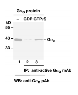
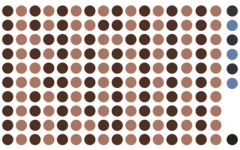
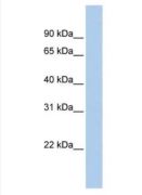
Example of Results
The following figure demonstrates typical results seen with Bioyears Biosciences Gαo Activation
Assay Kit. One should use the data below for reference only. 用户评论 产品评分 目前评分共0人 产品质量
售后服务
易用性
性价比
|
同品牌产品:
相关产品:
|


 m.cnreagent.com